You are here:Home » Solution » Featured Solutions » A Comprehensive Guide to Hantavirus Prevention and Control: Transmission Routes, Risks, and Laboratory Testing
A Comprehensive Guide to Hantavirus Prevention and Control: Transmission Routes, Risks, and Laboratory Testing
Views: 514 Author: Yammi Publish Time: 2026-05-14 Origin: Site
In recent years, Hantaviruses have continued to spread worldwide. Due to their high fatality rate and the risks of aerosol transmission and laboratory exposure, they have become one of the pathogens of primary concern in public health. With the continuous advancement of molecular diagnostic technologies, establishing safe and standardized molecular pathology laboratories has become increasingly important. Laboratory management and biosafety precautions are particularly critical during the detection of high-risk pathogens such as Hantaviruses. Next, we will explore the transmission routes of Hantavirus and the instruments and equipment commonly used in laboratory testing.
Classification of Hantaviruses
Infection with hantaviruses can lead to two types of severe diseases:
Hemorrhagic fever with renal syndrome (HFRS), also known as epidemic hemorrhagic fever, is the most common type of hantavirus infection. Typical clinical manifestations include fever, bleeding, and kidney damage. Severe cases may present with shock and multiple organ failure; without proper treatment, the case-fatality rate can exceed 10%.
Hantavirus Pulmonary Syndrome (HPS), which is primarily prevalent in the Americas, is characterized by pulmonary infiltrates and respiratory failure, and has an even higher case-fatality rate.
The prevalence of hantaviruses exhibits distinct seasonal patterns, with peak incidence typically occurring from November to January of the following year. In some regions, a minor peak may occur from May to July. The epidemic cycle is directly related to the activity patterns and population fluctuations of the rodent hosts.
Transmission Routes of Hantaviruses
Primary Transmission Routes
Rodents are the primary source of Hantavirus infection, with the brown rat and the black-striped field mouse being the most common hosts in China. The virus is shed through the host animal’s blood, saliva, urine, and feces. Humans are primarily infected through the following routes:
Respiratory Transmission: Inhalation of aerosols contaminated with excreta from infected rodents is the most common route of infection. The risk of exposure is significantly higher in enclosed, poorly ventilated spaces (such as warehouses, basements, and field shelters).
Gastrointestinal Transmission: Consuming food or water contaminated with excreta from infected rodents allows the virus to enter the human body through the mouth or the mucous membranes of the digestive tract.
Contact transmission: Infection can also result from bites or scratches by infected rodents, or from contact between broken skin or mucous membranes and the excreta or secretions of infected rodents.
It is important to emphasize that human-to-human transmission of Hantavirus is extremely rare. Clustered transmission within households has been reported only in a very small number of specific cases. Routine contact with confirmed patients does not lead to infection, so there is no need for excessive panic.
High-Risk Populations and Settings
The following groups are at significantly higher risk of Hantavirus infection than the general population and should take special precautions:
Residents of rural areas, particularly those engaged in agricultural production or fieldwork. This includes, but is not limited to, farmers, forestry workers, geological surveyors, and field construction workers.
Workers in farmers’ markets, grain warehouses, livestock farms, food processing plants, and warehousing and logistics facilities. Rodent activity is frequent in these environments, resulting in a higher probability of exposure to virus-contaminated materials.
Public health workers, medical laboratory technicians, and laboratory researchers. These individuals face occupational exposure risks when handling samples, conducting tests, or performing research.
Individuals with a recent history of camping, hiking, or staying in rural areas. This is particularly true for those who have visited forested or grassland areas with high rodent activity during the epidemic season.
Clinical Recognition and Prevention and Control Principles
Recognition of Early Symptoms
The incubation period for Hantavirus infection is typically 7–14 days, ranging from a minimum of 4 days to a maximum of 45 days. Early clinical manifestations resemble those of the common cold or influenza, which can easily lead to missed or misdiagnoses. High vigilance is warranted when the following symptoms are present and there is a relevant epidemiological history:
Fever: Sudden onset of high fever, typically above 38°C, which may be accompanied by chills and shivering; the fever usually lasts 3–7 days.
Systemic Toxic Symptoms: Pronounced “three pains”—headache, lower back pain, and orbital pain. These may be accompanied by generalized muscle aches, fatigue, nausea, vomiting, abdominal pain, and diarrhea.
Signs of capillary damage: The “three reds” are observed, characterized by flushing and redness of the skin on the face, neck, and upper chest. Conjunctival hyperemia and edema are present, and some patients may exhibit petechiae or ecchymoses on the skin and mucous membranes.
Currently, there are no specific antiviral drugs for Hantavirus. Clinical management primarily involves symptomatic and supportive care, with early intervention for high fever, hypotensive shock, and renal failure being the cornerstone of treatment.
Key Points for Daily Prevention and Control
The core of Hantavirus prevention and control lies in interrupting transmission routes, controlling sources of infection, and protecting susceptible populations. Specific prevention and control measures include:
Rodent Prevention and Eradication: Reducing rodent populations by improving environmental hygiene, eliminating rodent habitats, and using physical trapping devices or approved rodenticides is the most fundamental measure for Hantavirus prevention and control.
Food Safety Management: All food must be stored properly to prevent contamination by rodents. Drinking water must be boiled before consumption, and food that has been gnawed on by rodents or contaminated by their feces must not be eaten.
Personal Protection: When working or engaging in activities outdoors, avoid contact with rodents and their excrement as much as possible, and do not sit or lie on the grass. When necessary, wear a mask and gloves, dress in long-sleeved clothing, and promptly disinfect and bandage any broken skin.
Vaccination: Currently available inactivated Hantavirus vaccines provide over 90% protection against prevalent virus strains.
Key Points of Laboratory Testing
Laboratory testing is the cornerstone of Hantavirus diagnosis, and test results are critical for reducing the case fatality rate. Laboratory testing must strictly adhere to biosafety regulations. All procedures must be conducted in laboratories that meet biosafety level requirements.
1. Sample Collection and Processing
Common sample types used for Hantavirus testing include serum, plasma, whole blood, urine, and throat swabs. The appropriate sample types vary depending on the stage of the disease:
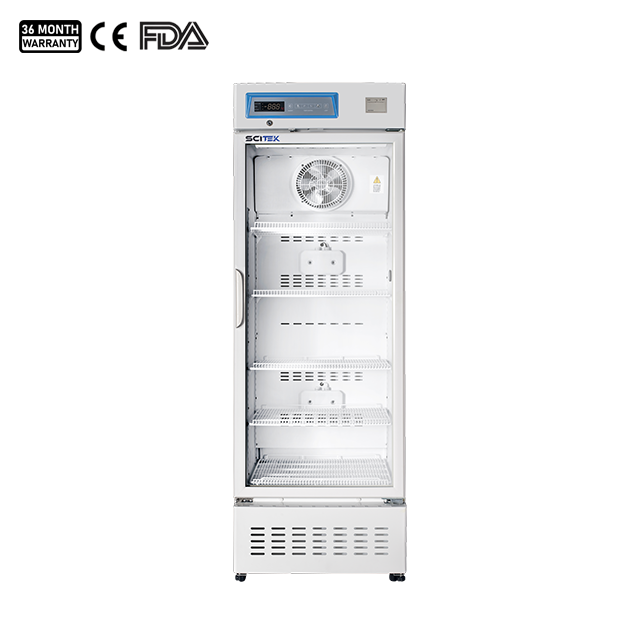
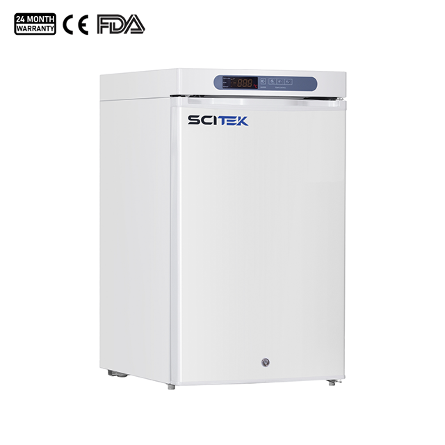
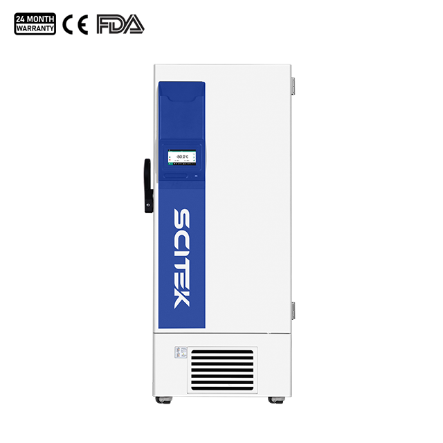
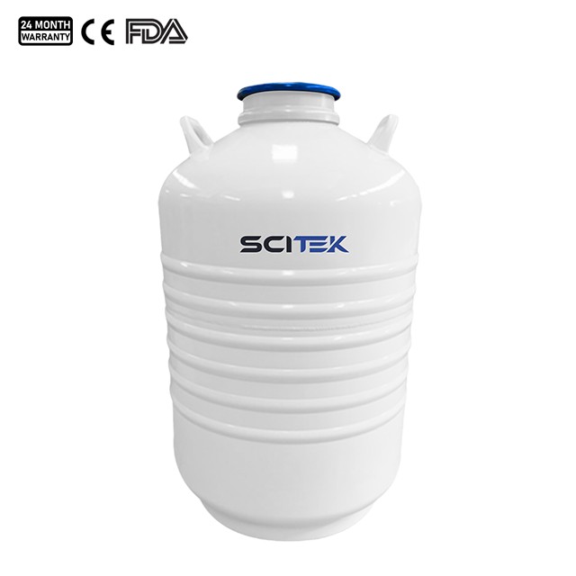
Acute-phase samples: Serum/plasma samples collected within 1 week of symptom onset. Suitable for nucleic acid testing, virus isolation, and IgM antibody testing. Samples should be submitted for testing within 48 hours at 2–8°C after collection. If long-term storage is required, samples must be stored at temperatures below –70°C to avoid repeated freeze-thaw cycles.
Convalescent-phase samples: Serum samples collected 2 weeks or more after onset of illness. Suitable for IgG antibody testing. A fourfold or greater increase in IgG antibody titers between paired serum samples may serve as a basis for confirmation of diagnosis.
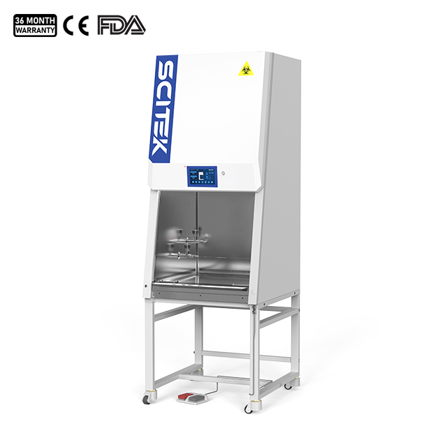
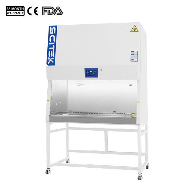
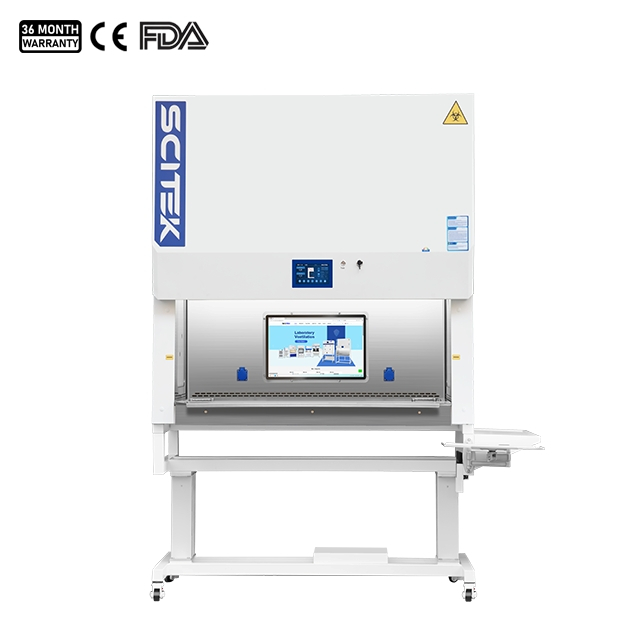
Strict personal protective measures must be followed during sample handling. All sample manipulation must be performed in a Class II biological safety cabinet to prevent aerosol generation. Instruments that have come into contact with samples must be disposed of in accordance with medical waste regulations to prevent cross-contamination.
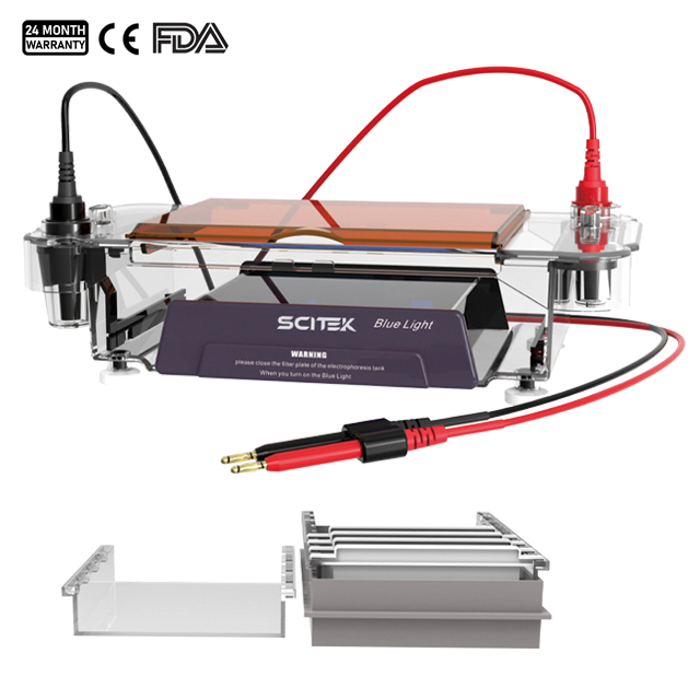
2. Common Diagnostic Methods
During an outbreak of hantavirus, diagnostic testing typically follows a stepwise process. From rapid screening of exposed individuals to confirmatory testing after hospitalization, the process generally proceeds in the following order.
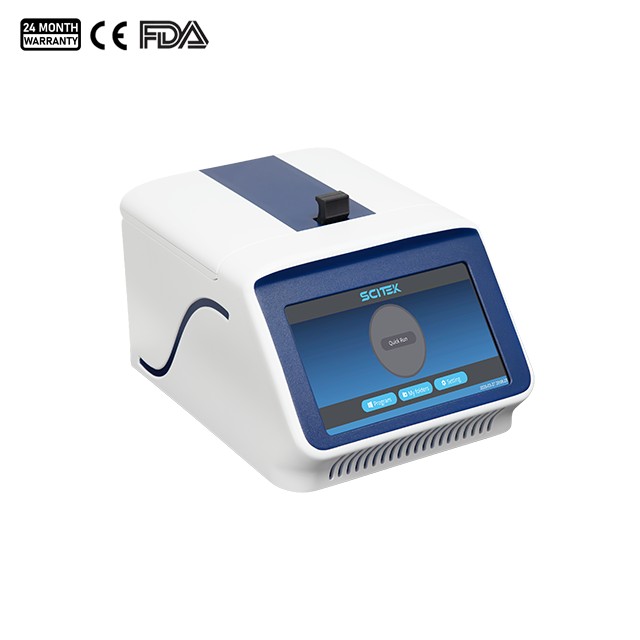
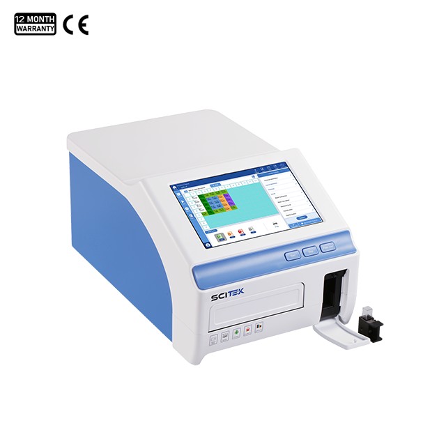
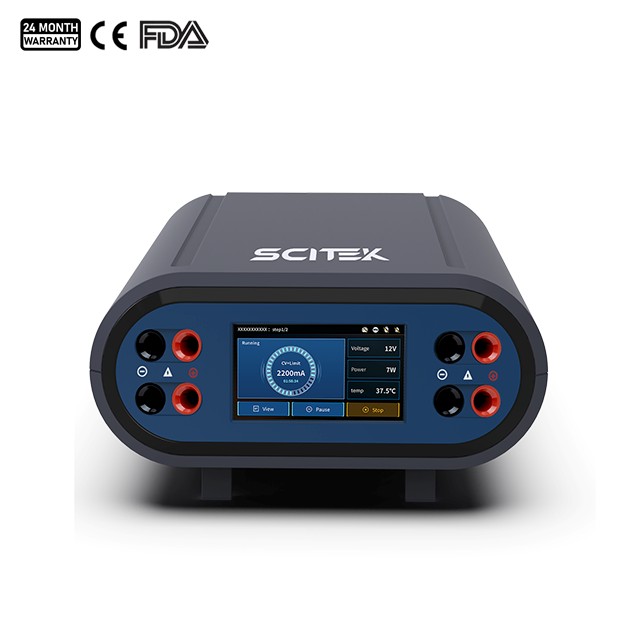
For patients in the early stages of infection, particularly within the first week after exposure, quantitative Real-time Fluorescence PCR Detection System (qRT-PCR) is widely used to directly detect Hantavirus RNA. In samples collected early in the course of the disease, the positive predictive value can exceed 90%.
qRT-PCR has the following characteristics:
High sensitivity
Simple operation
Low cost
Rapid test results
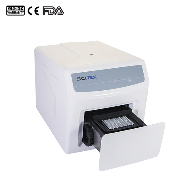
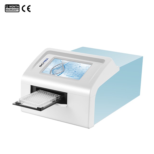
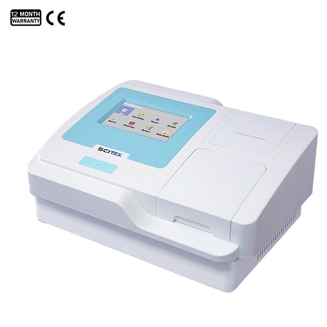
2. IgM Antibody Testing for Further Screening
For individuals with fever, a history of rodent exposure, or close contact with a confirmed case, rapid serological screening is typically the method of choice for further evaluation. IgM antibodies are usually detectable within 3 to 5 days after symptom onset, with a positive predictive value exceeding 95% during the first week of illness.
Common rapid screening methods include:
Gold particle method
Enzyme-linked immunosorbent assay (ELISA)
Immunofluorescence assay
The gold particle method is simple and rapid, making it suitable for emergency screening during outbreaks and in primary care settings.
3. Confirmatory Laboratory Diagnosis
Once suspected patients are admitted to a hospital or transferred to a specialized laboratory, more accurate confirmatory testing is performed.
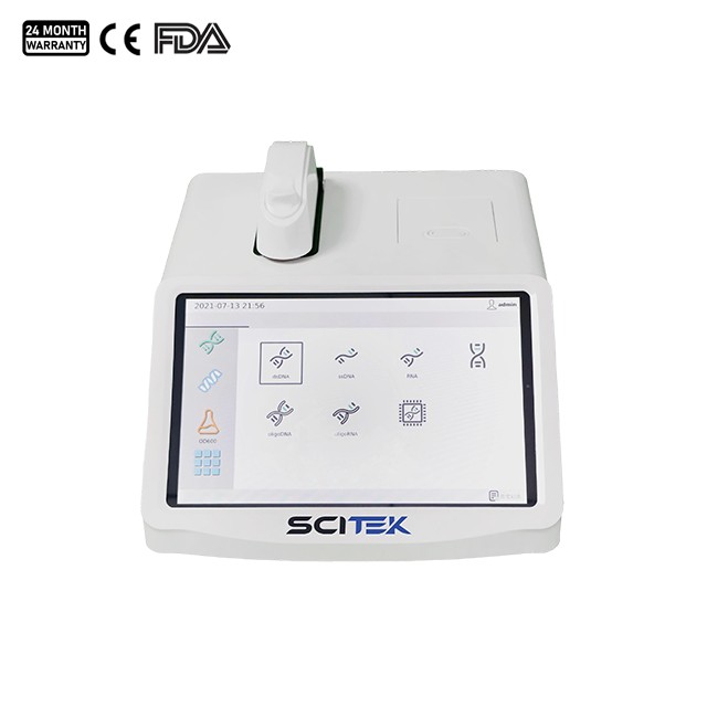
Serological antibody testing: This method offers higher accuracy
A positive IgM result typically indicates a recent or active infection.
IgG antibodies usually rise 1 to 2 weeks after the onset of symptoms and may persist for several years. A fourfold increase in IgG titers between acute-phase and convalescent-phase serum samples is considered strong evidence of active infection.
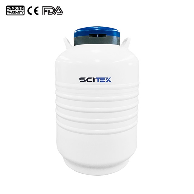
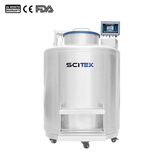
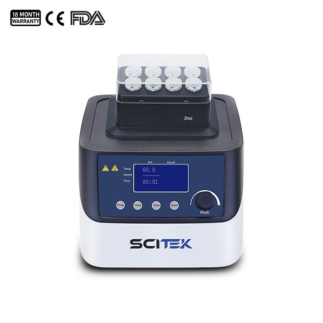
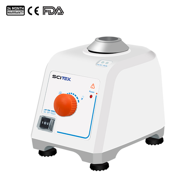
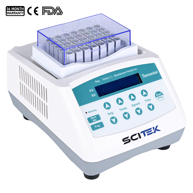
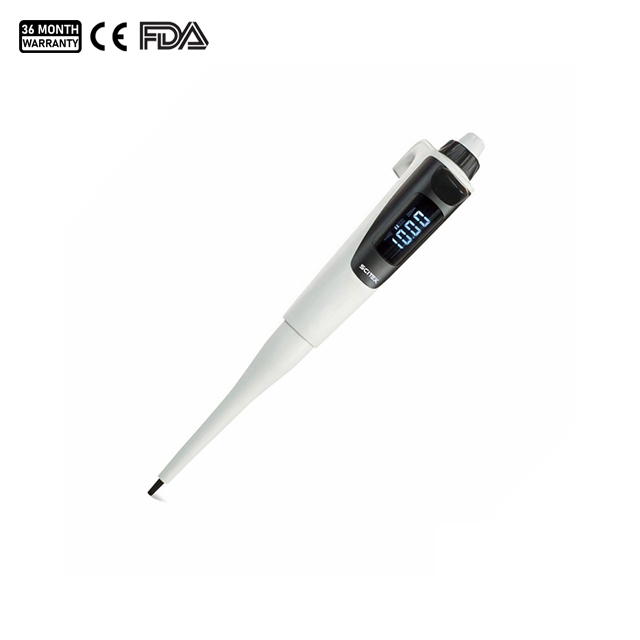
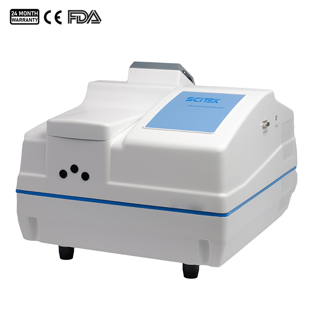
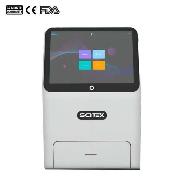
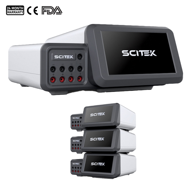
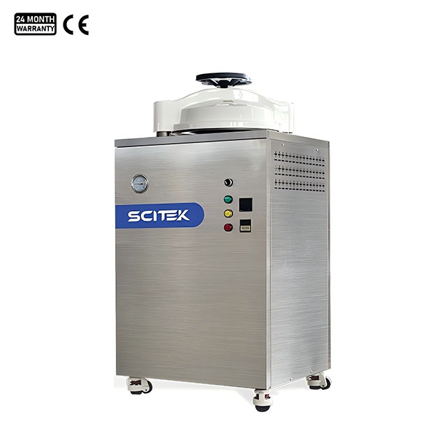
Laboratory Equipment Potentially Used for Hantavirus Testing
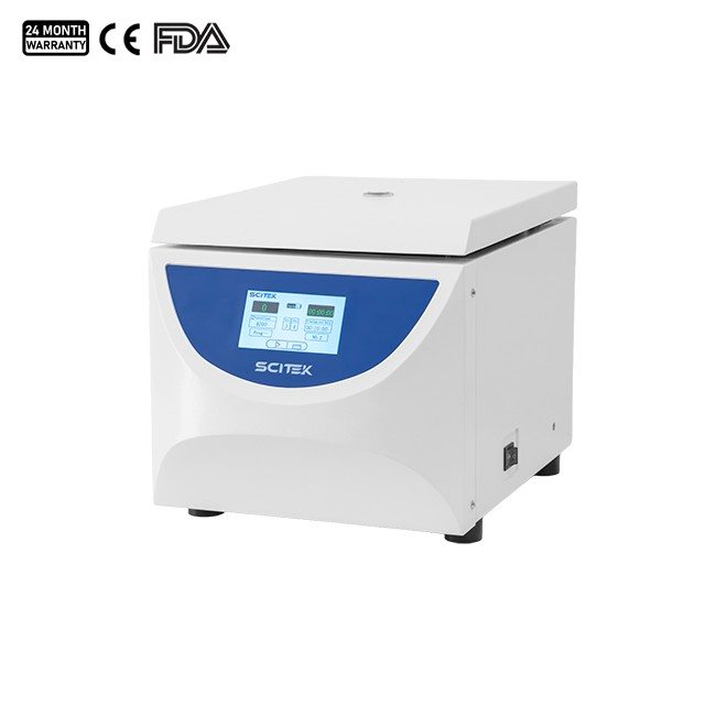
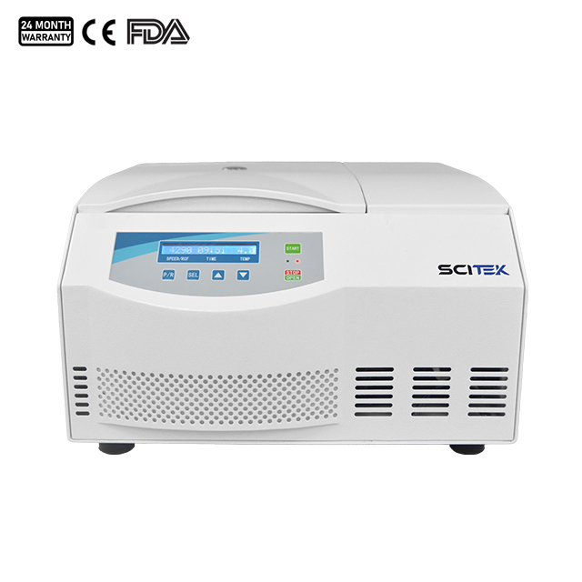
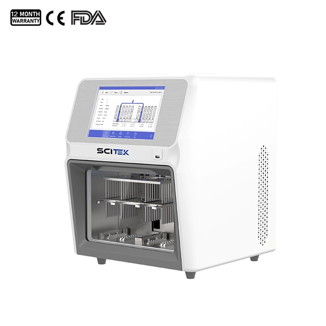
During virus testing, nucleic acid extraction, and sample processing, laboratories are typically equipped with the following devices.
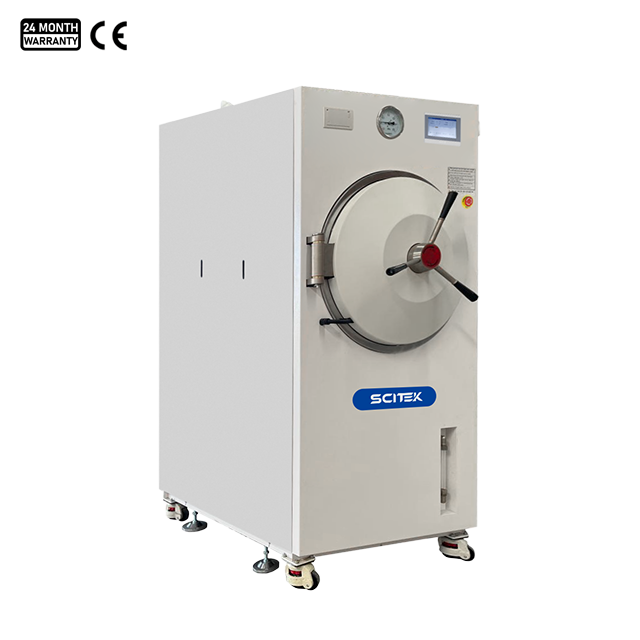
An essential component of the laboratory biosafety system.
Sterilization Temperature (℃) : 105~136
Max. Sterilization Pressure (Mpa) : 0.23
Drying Function : YES
Temperature (℃) : 121℃-134℃
Highest Working Pressure : 0.25Mpa
Temp.Accuracy : 0.5℃
Design Pressure : 0.28~0.3Mpa
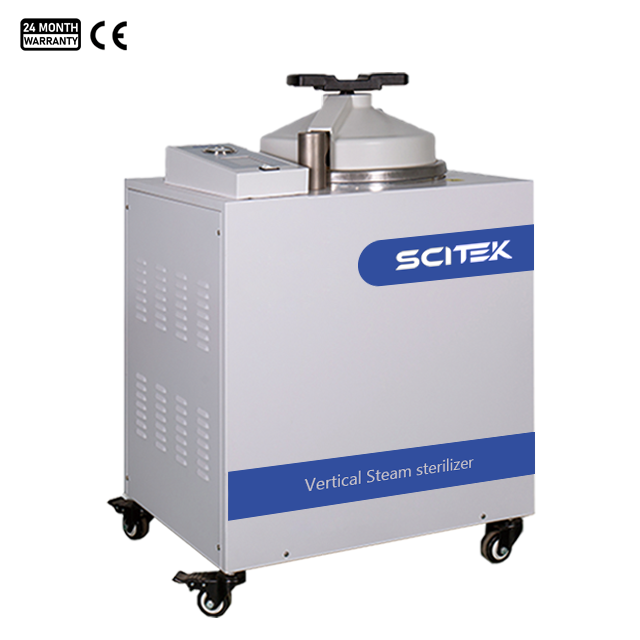
ST-VLA series with drying function
Sterilization Temperature (℃) : 105~136
Max. Sterilization Pressure (Mpa) : 0.23
Drying Function : YES
Water Injection Method : Built-in water tank
Drying Time : 15min
Conclusion
The accuracy of Hantavirus testing depends not only on advanced molecular diagnostic technologies but is also closely linked to laboratory biosafety management, sample handling procedures, and equipment configuration. Non-compliant operations, sample contamination, or inadequate equipment performance at any stage may lead to biased test results, thereby affecting disease diagnosis, outbreak surveillance, and clinical decision-making.
Therefore, establishing a comprehensive laboratory management system, equipping laboratories with reliable testing instruments, and strictly adhering to biosafety protocols are of critical importance for improving the accuracy of Hantavirus testing and ensuring the safety of laboratory personnel. With the continuous advancement of molecular diagnostic technologies, standardized, automated, and highly secure laboratory solutions will play an increasingly vital role in viral testing and public health prevention and control.
As a professional lab and medical equipment manufacturer, Scitek Global is certified by ISO 9001, ISO 13485, ISO 45001 and ISO 14001. Almost all our products are certified by ETL, CE and FDA .
Scitek Global looks forward to establishing cooperation with more distributors all over the world, and working together to create greater customer value.
 English