The microscope allows us to see many micro-organisms, cells, and organisms that the NAKED EYE cannot see. It enables scientists and researchers to observe the structure and form of cells, plants and animals, fungi, and viruses in greater detail. Microscopes are used not only in medical and scientific research but also in observing tiny circuits on circuit board chips, nanomaterials, geological samples, and particles or micro-organisms in food, among many other applications. Let's look at some of the various topics related to microscopes.
Types, Purposes of Microscopes
Different microscopes are used in other industries or places and have unique uses in different fields and experiments. Let's take a look at the features of various types of microscopes to help you better observe and study other aspects of the microscopic world.
| Types |
| Feature | Purposes |
| Optical Microscope | Biological Microscope | Pros:
1. Ideal for observing transparent biological samples, including cells and tissues, for biological or medical research.
2. Provides magnification from 4x - 100x for observation and analysis of cellular structures.
Cons:
1. Typically only suitable for transparent materials, larger tissue sections, or biological samples.
2. There are limitations to the observation of living cells. | Suitable for intermediate and elementary biological research, medicine, education, etc., such as observing the morphology and structure of cells, fungi, and bacteria.
|
|
| Inverted Microscope | Pros:
1. Lens facing up, suitable for long-term observation of liquid culture cells, live cell culture and cell behavior.
2. Allows observation of cells or the lower part of the liquid culture dish.
Cons:
1. Higher cost
2. Not suitable for general biological specimen observation | For long-term observation of cell cultures and live cell experiments |
Polarizing Microscope
| Pros:
1. Allows observation of optical properties and structure of materials.
2. Applies to crystallography, materials science and mineralogy.
Cons:
1. Requires specific sample and light source conditions.
2. Highly expensive and operators need to have specialized knowledge and skills. | Suitable for observing the structure, composition and optical properties of mineral or rock samples; structure, birefringence, polarising colors and defects in crystalline materials; fibrous proteins, cellular tissues and bone structures. |
Stereoscopic Microscope
| Pros:
1. Provides stereoscopic imaging for viewing opaque or three-dimensional samples.
2. Can use for large or irregular samples.
Cons:
1. Low magnification.
2. Limited observation of cells or microstructures. | Ideal for observing large samples such as insects, rocks, workpieces, and machine parts. |
| Electron Microscope | Scanning Electron Microscope (SEM) | Pros:
1. SEM provides extremely high resolution to show the minute structure and details of the surface of a substance, even in the nanometre range.
2. Can provide a three-dimensional image of the surface of a substance, which helps to observe the morphology, structure and composition of the surface of a substance.
3. Capable of observing larger and irregularly shaped samples without special preparation processes.
4. Different imaging modes, such as secondary and reflective electron imaging, can be used to obtain information about surfaces of other properties.
Cons:
1. Samples usually require fine processing and metal plating under vacuum for observation.
2. Equipment is expensive and running and maintenance costs are high.
3. Specialised training and skills are required | Suitable for studying the microstructure and composition of materials such as metals, ceramics, rocks, minerals, soils, polymers, insects, tissues and nanomaterials.
|
| Transmission Electron Microscopy (TEM) | Pros:
1. The TEM provides exceptionally high resolution, enabling observation of the internal structure and composition of samples at the atomic and molecular levels.
2. An energy-dispersive X-ray spectroscopy (EDS) accessory enables elemental analysis of samples.
3. The TEM can handle relatively large sample sizes.
Cons:
1. Samples need to be prepared in fragile sections using complex techniques such as ion thin sectioning or cryosectioning.
2. Expensive equipment with high maintenance and running costs
3. Specialised skills of the main operator
4. Operates under vacuum | Suitable for studying molecular or crystalline defects and structures in rocks, minerals, soils, metals, ceramics, polymeric materials and nanomaterials. |
| Fluorescence Microscope | Fluorescence Microscope
| Pros:
1. Can fluorescently label specific biomolecules
2. Enables more intuitive observation of the location and distribution of specific molecules in cells and tissues.
3. Supports observation of cell metabolism, division, apoptosis, etc.
Cons:
1. Requires specific fluorescent dyes or labelling.
2. Microscopes are more costly and require specialised handling. | It belongs to a kind of advanced microscope, which is widely used for observing fluorescently labeled biomolecules, such as proteins, DNA, RNA, etc. |
| Other | Compound microscope
| Pros:
1. Ideal for teaching and beginners.
2. Simple and easy to operate.
Cons: 1. Low magnification. 2. Limited functionality and specialisation | Simple microscope suitable for teaching purposes, easy to use and operate. Often equipped with LED illumination, suitable for student learning and beginners. |
Digital Microscope
| Pros:
1. Can be connected to a computer or display for real-time image display and storage
2. Images can be easily saved, shared and analyzed, often with digital image capture and processing capabilities
Cons:
1. Depending on the computer system, it may have connection or compatibility issues.
2. Typically, it magnifies at 4x - 100x magnification, which is unsuitable for high magnification needs. | Education, industrial inspection, and medical diagnosis. For real-time observation, image analysis, distance learning and remote collaboration. |
| Near Field Scanning Microscope (AFM/STM) | Pros:
1. Provides very high resolution, enabling the observation of surfaces of matter at the atomic scale
2. AFM is suitable for observing surface morphology and mechanical properties, while STM is used for observing surface atomic structure and electrical conductivity.
3. No special sample preparation is required before observation
4. Provides information on the surface morphology, electronic structure, and mechanical properties of the sample.
Cons:
1. Higher skill requirements for operators
2. Expensive equipment
3. Longer imaging time | Ideal for observing the morphology and mechanical properties of nanomaterials as well as biomolecules |
|
Buying the right microscope: factors to consider
Microscopes have different applications and uses. When choosing one, you need to consider the type of sample to be observed, the magnification, the budget and the purpose.
Type of sample:
The type of microscope can be quickly selected based on the type of sample to be observed:
| Type | Compound microscope | Stereomicroscope |
| Magnifying power | 40x-1600x | 5x-160x |
Viewing mode
| Provides a flat image that the observer views in the eyepiece | Provides a three-dimensional view where the observer can obtain a three-dimensional perspective by looking through both eyepieces simultaneously. |
| Sample type | Suitable for the study of microscopic organisms and cells, e.g., bacteria, pond scum, blood samples and aquatic organisms (samples need to be processed, prepared in thin sections or stained) | Study of larger specimens such as beetles, bugs, gemstones, soils, and small plants and animals (usually without processing the sample) |
Recommendations based on experience:
1. For handling environmental samples such as soil, rocks, etc., choose a dark-field microscope.
2. For blood samples, choose a phase contrast microscope.
3. For clinical or medical samples such as protein, DNA, etc., choose a fluorescence microscope. 4. for large samples, choose stereomicroscope.
4. For large samples, choose stereomicroscope.
Resolution and magnification:
Resolution: depends on the level of detail of the sample you need to view. Higher-resolution microscopes can demonstrate finer cellular structures or microscopic tissues.
The objective and eyepiece of the microscope determine the magnification, and multiplying the values of the two factors gives the total magnification of the microscope. Depending on the size and fine structure of the sample, you need to observe, choose the appropriate magnification.
Typically, compound microscopes have four objectives: 4x, 10x, 40x, and 100x; however, some also have five objectives.
Microscopes usually have three types of eyepieces: monocular, binocular and trinocular.
Light source and contrast:
LED (Portable Microscope): Produces a bright, cool light that does not generate heat and is usually fitted with a dimmer and characterized by low price and long life. Use of batteries also supports outdoor use of the microscope.
Halogen Lamp: Produces strong white light, generates heat quickly, is usually equipped with a dimmer, and is the standard light source in many pedestal microscopes. Lifespan is moderate.
Tungsten/Incandescent: produces a warm white light and generates heat quickly, usually not equipped with a dimmer system, often used in beginner microscopes. Short-lived and inexpensive.
Fluorescent: Produces white light, produces less heat, usually used in professional microscopes. Short life span.
Contrast: If the microscope is provided with a contrast adjustment function, it can enhance the details of the image and make the observation more transparent.
Illumination Methods:
Microscopes are illuminated in various ways, with different types of illumination suitable for different types of samples and observation needs. How a microscope is illuminated is closely related to the type of light used and the settings.
The following are a few common types of microscope illumination:
Brightfield illumination: For transparent samples, such as biological samples or cells. An incandescent or white LED lamp is required to produce the right amount of light.
Darkfield illumination: Suitable for opaque samples such as bacteria or colorless crystals. A circular light source or light fixture is required.
Phase Contrast Microscopy: For opaque samples. A typical light source is a ring light source or a unique optical device that produces a clear image by enhancing the phase difference.
Fluorescence Illumination: Suitable for the observation of fluorescently labeled cells or biomolecules. Typical light sources are mercury lamps, sodium lamps or LEDs.
Polarized Illumination: Suitable for viewing samples with anisotropic properties, such as crystal structures or fibers. The direction of vibration of the light source needs to match the demand of the polarising filter to produce polarised light.
Operating Difficulty:
Beginners can choose microscopes that are easy to operate and easy to maintain. You can select the additional functions of the microscope according to your needs, such as autofocus, video camera, etc.
Common additional accessories:
Camera: the camera is mounted on the objective lens and lets you take pictures of the analyzed sample.
Screen: It allows you to display your observations under the microscope in real-time for easy compliance.
Discussion Bridge: This tool allows the beam to be separated and displayed in two different objectives so that two users can observe the sample simultaneously.
Slide Scanner: Allows scanning the observed sample and processing the collected images through the computer.
Zoom: The zoom allows continuous magnification of the observed sample.
Motorized system: For moving the nosepiece.
Quality:
You can check the quality of a microscope by looking at its light source, construction, and materials, from cheap plastic to durable, corrosion-resistant stainless steel metal body.
After-sales service:
Choose a company that offers comprehensive after-sales service and support to ensure ease of maintenance and repair of the equipment.
Components and construction of a microscope

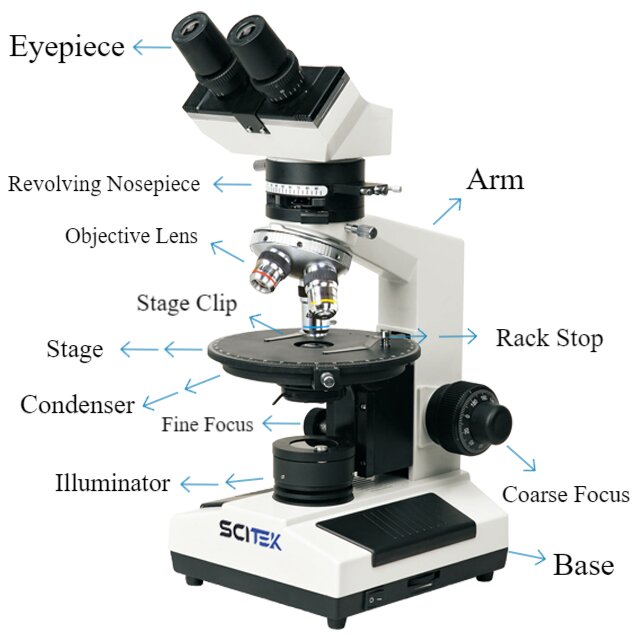
Eyepiece/Ocular:
The eyepieces are located at the top of the optical path and the top of the barrel, usually two. Eyepieces are often interchangeable and can vary in magnification. The eyepieces typically have a fixed magnification, e.g. 10x.
Objective Lens:
Objective lenses located at the bottom of the microscope are used to magnify and focus the sample. Often, a microscope has several objectives with different magnifications, e.g., 4x, 10x, 40x, 100x, etc.
Low Power Objectives: 4x or 10x magnification for quick orientation and initial observation.
High Power Objectives: 40x or 100x magnification for high magnification observation of cells and microstructures.
Oil Immersion Objectives: Typically 100x magnification, requiring oil mirror oil to improve resolution.
Stage:
Used for placing samples to be observed. It may include clamps or mechanical stages to ensure specimen fixation. Generally, it can be moved vertically or horizontally to position the specimen for examination.
Condenser:
Situated beneath the stage, the condenser is pivotal in directing light onto the specimen. Comprised of multiple lenses, its primary function is to concentrate and align the light source, ensuring even specimen illumination. Typically, the condenser can be modified regarding height and aperture size, enabling control over light intensity and angle.
Illumination Source
It provides illumination and ensures light passes through the sample and into the objective lens.
Diaphragm/Iris
We are used to adjust the intensity and contrast of light and to control light flux.
Focus Knobs:
Knobs or handles are used to adjust the focusing adjustment device manually. We are used to adjusting the focus between the objective lens and the sample to ensure a clear image.
Coarse Focus: For fast focus adjustment.
Fine Focus: Used for a small range of focus adjustments to achieve finer focus.
Body Tube
The body tube serves as the link between the eyepiece and the objective lenses. Housing a set of lenses, it works to amplify and transfer the image from the objective to the eyepiece. Certain microscopes allow adjustment of the body tube, enabling extension or tilting for user comfort and ergonomic positioning.
Arm
The overall support structure of the microscope protects the optical components and provides support.
Base
The microscope's foundation, or base, functions as a stable platform supporting the entire instrument. It commonly houses the illumination source, power switch, and electrical controls. Its primary role is to ensure the stability of the microscope while in use.
Guide to Using a Light Microscope:
Step 1: Connect the light microscope to a power source, unless natural light via a mirror is available.
Step 2: Rotate the revolving nosepiece to align the lowest magnification objective lens.
Step 3: Prepare the specimen by placing a coverslip for protection before mounting it on the stage.
Step 4: Secure the slide with the metal clips, ensuring the specimen is centered beneath the lowest magnification objective lens.
Step 5: Look through the eyepiece and use the coarse adjustment knob to gradually bring the specimen into focus. Be cautious to prevent the slide from contacting the lens.
Step 6: Adjust the condenser for optimal light intensity. Modify the diaphragm under the stage if needed, especially with low-power objectives.
Step 7: Refine the focus by gently using the fine adjustment knob until a clear image of the specimen is achieved.
Step 8: Thoroughly observe and analyze your specimen.
Step 9: After using the lowest power objective, switch to the medium power objective and readjust focus using the fine adjustment knob.
Step 10: Once focused, move to the high-power objective for further detailed examination.
Applications of microscopes
Medicine:
A microscope is an important auxiliary instrument for laboratory professionals or researchers to study cells, DNA, bacteria, viruses, pathological sections, etc. It helps one better understand the fine structure and characteristics of microorganisms. It contributes immensely to finding treatment options for various diseases.
Materials and Nanotechnology:
The microscope can observe the structure and appearance of materials at the nanoscale, which promotes the development and progress of nanotechnology. It can also follow the crystal structure of metals or alloys, which helps to improve the properties of materials.
Education:
We all see microscopes in school labs where students learn about the miniature world through microscopes. Students, through the microscope, can directly view plant organs or cells and many other tiny structures not visible to the NAKED EYE, such as chloroplasts and platelets. Microscopes promote students' knowledge and understanding of the microscopic world.
Biology:
Microscopes play a vital role in studying genetics, physiology and ecology. It is often also used to help police identify trace evidence left at crime scenes, such as fingerprints and hair.
Geology:
Microscopes allow observation of the microstructure of soils, rocks, and minerals, which helps to study the type of geology and the evolution of the environment. It is essential for the stability of ecosystems and the protection of the environment.
Conclusion
As one of the important instruments in the laboratory, microscopes are essential for many experimental workflows and analyses. Microscopes are also more advanced nowadays to meet the needs of many specific applications.
Scitek has a wide range of high-quality microscopes, including fluorescence, compound and benchtop binocular stereo microscopes. We have our manufacturing plant and welcome visits from all over the world.
Top Questions
What is a microscope?
Simply put, a microscope is an optical instrument that can magnify tiny objects dozens to hundreds of thousands of times. It allows cells or samples not visible to the naked eye to be magnified indefinitely through a lens or objective lens, thus revealing many small details that cannot be seen.
What does "microscope" mean?
The word "microscope" comes from the Greek words "mikros," meaning "small," and "skopein," meaning "to look at." It has been used in English since the early 17th century.
Who invented the microscope?
The invention of the microscope has a relatively long history and is still being determined who invented it. The earliest microscopes date back to the early 17th century.
The first compound microscope was invented around 1590 by Zacharias Janssen and his father Hans Janssen.
How much is a microscope
The price of a microscope can vary depending on various factors; in general, basic and entry-level microscopes designed for educational purposes or hobbyists can range from about $50 to $500.
More advanced microscopes, such as compound microscopes with higher magnification and high-quality optics, can range from $500 to $2000. These can be used in laboratories, for more in-depth educational purposes, or by hobbyists looking for higher-quality imaging.
Highly specialized or professional microscopes, including those used for scientific research, medical laboratories, or industrial applications, can range from $2,000 to hundreds of thousands.

 English