The microscopic world of life is full of infinite mysteries and explorations, and microbiology is one of those fascinating areas. Whether in food safety, environmental monitoring or scientific research, we need accurate and efficient tools to reveal the presence and number of microorganisms.
In modern microbiology research and laboratories, the colony counter is an indispensable piece of equipment in microbiology laboratories. It can count and analyze microbial colonies quickly and accurately, providing reliable data support for scientific research. Fully automated colony counters are an efficient tool that has emerged recently.
In this article, we will introduce various common knowledge about colony counters and analyze their key performance indicators in detail.
Development of colony counting methods
As a part of microbiology and laboratory technology, colony counters have undergone several stages of development and change. From the earliest manual counting to the application of modern automated technology. The following are the major historical changes in the development of colony counters:
1. Manual counting stage: The earliest colony counting method was to manually count microbial colonies on agar plates by visual observation. This method is simple, but the workload is large and prone to error.

2. Introduction of counting chambers: At the end of the 19th century, special chambers (Petri smear and counting compartments) were introduced. These devices helped researchers to count microbial colonies more accurately. At the same time, the technical chambers helped reduce the environmental impact and improved the accuracy of the counts.

3. Electronic counters: At the beginning of the 20th century, with the development of electronic technology, electronic counters were gradually applied to colony counting. These devices use electronic technology for counting, improving the speed and accuracy of counting.
4. Automation and digitalization: With the advancement of computer technology, colony counters have gradually become automated and digitalized. Counting and data processing has become more efficient and accurate, while more detailed analysis reports can be generated.
5. Image analysis technology: In the 21st century, image analysis technology has been applied in colony counting. Digital cameras and image processing software can capture and analyze images of colonies on agar plates to automate counting and analysis.
6. Real-time monitoring technology: Real-time monitoring of microbial growth and colony formation combines sensors, automated systems, and real-time data transmission to monitor microbial growth more promptly.
7. Mmicrofluidic technology: With the support of microfluidic technology, colony counting, and analysis become more miniaturized and have high throughput. Microfluidic chips allow for simultaneous counting and analysis of multiple colonies on a tiny scale.
How to count bacterial colonies
Direct observation
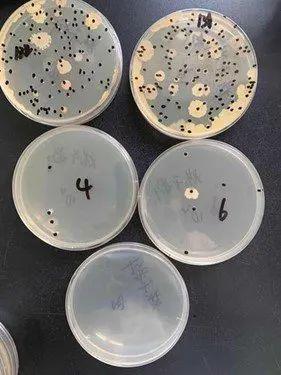
All visible colonies are counted by visual observation. Although most of the daily tests are done by pouring plates, the smear method is more advantageous in detecting heat-sensitive organisms. It gives better counting results and is more suitable for strictly aerobic bacteria. The disadvantages of the smear method are non-drying of the agar surface during smearing and spreading colonies after incubation failing to count.
Colony counter
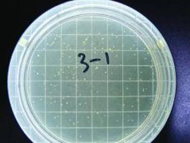
The number of colonies on a plate can be counted by direct visual observation or with the help of an instrument. The colony counter is a digital display type automatic bacterial testing instrument. It comprises a counter, probe, counting pool, and other parts.
The working principle of the electronic counter is to determine the resistance change of the liquid in the small hole. The small hole can only pass through a cell; when a cell passes through this small hole, the resistance increases significantly, forming a pulse automatically recorded on the electronic recording device. The method is more accurate in its measurements, but it only identifies the particle size and does not distinguish whether it is a bacterium. Therefore, the bacterial suspension must not contain any debris.
A usual colony counter is divided into three types: manual, semi-automatic, and fully automatic, and different counters can be selected according to the requirements for accuracy.

3. Turbidimetric method
The turbidimetric method indirectly determines the number of bacteria according to the light transmission of bacterial suspension. The concentration of bacterial suspension in a certain range and the transmittance are inversely proportional to the optical density. Therefore, a photoelectric colorimeter can be used to determine the bacterial suspension, and the optical density (OD value) indicates the concentration of the sample bacterial suspension.
This method is simple and fast, but it can only detect the suspension containing a large number of bacteria; the relative number of bacteria and the color of the sample are too dark and can not be determined by this method.
4. Determination of total cellular nitrogen or total carbon
Nitrogen and carbon are the main components of the cell; the content is more stable, and the Determination of nitrogen and carbon content can be deduced from the quality of the cell. This method is suitable for higher concentrations of microbial detection.
5. Live cell counting method
Commonly used plate colony counting method is based on the principle that each live bacteria can grow a colony design. Take a certain volume of bacterial suspension, make a series of dilutions, and then the quantitative dilution of plate culture, according to the number of cultured colonies, the number of live bacteria can be calculated.
This method is highly sensitive and detects the number of contaminated live bacteria, but also the current international microbiological detection methods used in many countries. The use of this method should be noted:
Generally select the number of colonies between the plate for counting in the 30 to 300. Too much or too little are inaccurate;
In order to prevent colonies from spreading and affecting the count, 0.001% of 2,3,5-triphenyl tetrazolium chloride (TTC) can be added to the culture medium;
This method is limited to colony-forming microorganisms. Widely used in water, milk, food, medicines, and other materials, bacterial testing is a commonly used microbiological detection method.
6. Determination of cell weight method
This method is divided into wet and dry weight methods. Wet weight method is a unit volume of culture by centrifugation after the wet bacterial body for weighing; dry weight method is a unit volume of culture by centrifugation, wash with water and put into the desiccator heating and drying, so that it loses moisture and then weighing. This method is suitable for samples with high concentrations of bacteria and is a common method for microbiological testing.
How to count bacterial colonies on agar plate
According to the principles of ISO 7218: 2007 (E) colony counting, the counting was performed as follows:
If there are two consecutive dilutions of the plate colony number (N) in the range of 100 ~ 300, according to the formula below.
N = ΣC/Vx1.1 xd : N a plate colony number; ΣC - plate (including the appropriate range of colony number of the plate) the sum of the number of colonies; V - inoculation of the volume of bacterial solution; d - dilution factor (the first dilution).
The results are reported in whole numbers with two significant figures, trimmed to the nearest whole number according to the principle of "rounding". They can also be expressed in the form of an exponent of 10 (scientific notation), trimmed to two significant figures according to the principle of "rounding".
When the number on the plate is less than 10, it is counted in four cases. 1:
When the number of colonies on the plate is less than 10 and greater than 4, the results are calculated using the methods in Part I and reported as estimates.
When the number of colonies on the plate is less than 3 and greater than 1, the precision is too low and the results are reported as microorganisms detected and less than 4/d CFU/g(mL).
When there are no colonies on the plate, the result is less than 1/d CFU/g (or CFU/mL), d is the dilution factor, d=100=1 when the sample is not diluted.
If the number of colonies in the first dilution d1 is greater than 300 and all are typical or confirmed colonies, the number of colonies in consecutive dilution d2 plates is less than 300 and there are no visible typical or confirmed colonies. The counting method was as follows:
Results are recorded as less than 1/d1CFU/g (or CFU/mL) and more than 1/d2CFU/g (or CFU/mlL) .
If the number of colonies on the plate at the first dilution d1 is greater than 300 and there are no typical or confirmed colonies. The counting method is as follows:
The result is recorded as less than 1/d2 CFU/g (or CFU/mL).
When the first dilution d1 plate has a colony count greater than 300, the second consecutive dilution d2 plate has a colony count less than 10.
If the plate colony count for the first dilution d1 is between 300 and 334, count as before.
If the plate colony count for the first dilution d1 is greater than 334 and the plate colony count for the second consecutive dilution d2 is greater than 8 and less than 10, the count is reported as an estimate and is less than 1/d2 CFU/g (or CFU/mL).
If the plate colony count for the first dilution d1 is greater than 334 and the plate colony count for the second dilution d2 is less than 8, the result is invalid.
Plate colony counts on two consecutive dilutions d1 and d2 are greater than 300 and results are reported as follows: greater than 300/d2 or greater than 300 x b/A x 1/d2. b is the number of positive colonies confirmed in suspect colony A.
When the number of colonies (N') in the plate at the highest dilution is between 10 and 300, calculate according to the following formula:
N'=c/V×d where: c a number of colonies on the plate; V a volume number of inoculated bacteria; d - dilution factor.
When there is a spreading colony, if the spreading colony is less than one-fourth of the plate area, the number of colonies in the remaining part can be calculated, and then the number of colonies in the whole plate can be deduced; if it is greater than one-fourth, it cannot be counted; a chain colony, only as a colony count.
Some requirements in the determination of colony counts
In order to correctly reflect the presence of various aerobic and partially anaerobic bacteria in food, some of the following requirements and regulations must be followed in the test:
Utensils and diluents used:
Glassware used in the test, such as Petri dishes, pipettes, test tubes, etc., must be fully sterilized and thoroughly washed before sterilization, with no residual bacteriostatic substances.
The liquid used as sample dilution should have a blank control for each batch. If several colonies appear on the agar control plate, additional control plate. In order to determine the blank dilution, used to pour the medium of the petri dish, or petri dish pipette or air pollution may exist.
Check the dilution of the sample. If appropriate, it can be sterilized or distilled water, but use phosphate-buffered saline, especially 0.1% peptone water. Because peptone water on the bacterial cells has better protection, not because the process of dilution and food samples have been damaged in the original bacterial cells, leading to death.
Dilution of test samples:
Ensure the container is sterile when diluting the test sample.
When removing the sterilized pipette from the tube, do not touch the tip of the pipette to the exposed part of the other pipettes still in the container, and do not touch the outside of the mouth of the bottle and the mouth of the test tube when entering or exiting glass bottles and test tubes containing diluted liquids;
When inhaling the liquid, it should first be above the pipette scale. Then lift the pipette tip away from the liquid surface and put the tip against the inner wall of the glass bottle or test tube to make suction.
Plate inoculation and culture:
When adding the dilution solution to the sterilized petri dish, select 2 to 3 suitable dilutions, respectively; for 10-fold incremental dilution, each dilution should be made of 2 Petri dishes.
Nutrient agar for pouring Petri dishes should be pre-heated to melt and kept warm at 45 ± 1 ℃ in a constant temperature water bath to be used. When pouring Petri dishes, each dish into about 15mL, and finally, the bottom of the agar with precipitation part of the discard.
To prevent bacterial proliferation and the production of flaky colonies, the test solution should be added to the dish within 20 minutes after adding agar into the dish, and immediately make it and the agar mixed evenly.
When mixing the test sample with the agar, the bottom of the dish can be rotated on a flat surface in one direction first and then in the opposite direction in order to make full mixing. Automatic mixer can also be selected.
Do not leave the agar in the dish long after it solidifies. The agar should be turned over and incubated within a few minutes after solidification to avoid spreading of colonies. If necessary, the dish can be opened and inverted (bottom of the dish upwards) in the incubator after 15-60 minutes to make the surface of the agar dry, and then move the bottom of the dish to the cover and invert it in the incubator for cultivation.
Control tests:
To avoid confusion with bacterial colonies, a petri dish of test dilution mixed with agar may be left at 4°C without incubation to be used as a control when counting pick-up colonies.
TTC solution should be kept in a cold, dark place to prevent decomposition by heat and light.
Colony counting:
When removing the Petri dishes from the incubator for colony counting, the growth of colonies on the plates should first be observed on two Petri dishes of the same dilution and several containers of different dilutions, respectively. The number of settlements should be close to the number of 2 plates in the parallel test. The number of colonies on several plates at different dilutions should be inversely proportional to the dilution of the test sample, i.e., the greater the dilution of the test sample, the lower the number of colonies. The smaller the dilution, the higher the number of colonies.
the number of colonies should be selected between 30~300 plates as the standard for colony counting. 1 dilution using two plates, the average of the two plates should be used; such as one of the plates has a larger piece of colony growth, it is not appropriate to use
Precautions
If the number of colonies on the plate with a large dilution is higher than those with a small dilution, it is an error in the test work, a laboratory accident. In addition, this may also be due to bacteriostatic agents mixed into the sample, which can not be used as the basis of the sample count report.
If the plate appears to have chain-like colonies, there is no obvious boundary between the colonies. This is caused by bacterial mass dispersing when the agar is mixed with the test sample.

CCD Sensor
The CCD sensor is one of the most critical components in a colony counter. Its grade can seriously affect the stability of the counting and analysis results. Today's imaging hardware includes photo imaging and scanning imaging.
Photo imaging: fast imaging speed can ensure that the colony image is in 0.5 seconds, but also can be multi-point or single-point autofocus, and pixel resolution is generally higher. However, the disadvantage is that the light intensity in the imaging environment can not be accurately identified to the center of the image and the edge of the image to maintain full consistency; to a certain extent, it interferes with the automatic identification of the colony target.
Scanning imaging: to solve the problem of uneven brightness of the colony target, counting and analysis of stable, simple operation and fast imaging speed, adjustable imaging resolution. The disadvantage is expensive.
Image acquisition channel
Image acquisition channel is another important part of the colony counter. It determines the acquisition speed and stability of the colony image. Choosing a counter with high speed, stability, and many acquisition channel indicators can improve experimental efficiency and accuracy.
Colony resolution
Colony resolution refers to the smallest colony size the counter can distinguish and count. Higher colony resolution means the counter can recognize smaller colonies, thus providing more accurate counting results.
Typical entry-level colony counters have a minimum resolution of around 0.1mm.
Ease of use and functionality of the device
Choosing a colony counter that is easy to operate and has the necessary functions can improve the efficiency of the experiment. For example, touch screens, automatic counting and data storage and export, data analysis and report generation, strain library management, and other functions can simplify the operation process and improve data management efficiency.
Analysis software
In the analysis, software selection should be considered for all types of imaging interference with the automatic discharge ability. It is best to choose analysis software with an automatic correction function in order to improve its imaging effect automatically.
Convenience
Convenience is also one of the factors to consider when choosing a colony counter.
Price and after-sales service
Finally, you should also consider the price and after-sales service of the colony counter. The price may vary depending on the brand, model, and features, so choose according to your lab's budget. Knowing about the supplier's after-sales service and technical support is important to get timely help and support when needed.

 English


